If you’ve ever watched a patient roll out of a hospital examination room clutching a crumpled ECG printout, or seen a cardiac nurse quickly press a button to capture a 10-second snapshot of a heart’s electrical activity, you’ve probably interacted with the workhorse of cardiology: the 6-channel ECG machine. For 15 years, I’ve been supplying these devices to clinics, private practices, and urgent care centers across the Midwest, and the question I get most often isn’t about how to turn the machine on—it’s about how it handles the messy, diverse rhythms that show up on the screen. At first, I’d ramble through technical specs that left clinic managers glassy-eyed. Now, I break it down like I would over a cup of coffee, because every rhythm a 6-channel machine processes isn’t just a line on a graph—it’s a story of a heart working (or not working) as it should. 6 Channel ECG Machine

Let’s start with the basics, because if you don’t get how a heart’s electricity works, you can’t wrap your head around how a 6-channel machine translates that into something usable. The heart’s electrical system starts in the sinoatrial (SA) node, a tiny cluster of cells in the right atrium that acts as the natural pacemaker. Each electrical signal travels down a specific path: from the atria to the atrioventricular (AV) node, through the bundle of His, and out to the ventricles. A standard ECG measures this activity in 12 different “leads”—pairs of electrodes placed on the arms, legs, and chest that pick up the signal from every angle. A 6-channel ECG machine doesn’t show all 12 leads at once, but it’s not missing critical information: it prioritizes the leads that capture the most common, clinically useful signals, and processes each rhythm to make those leads consistent and readable, even when the heart is misfiring.
The most common rhythm we handle is sinus rhythm, the “normal” baseline that every clinician knows. For a sinus rhythm, the SA node is firing at a steady 60 to 100 beats per minute (BPM). When a 6-channel machine processes this, it’s not just printing a flat line—it’s calibrating the amplitude (the height of each wave) so that the P wave (the atria contracting), QRS complex (the ventricles contracting), and T wave (the ventricles resetting) are clearly visible across all six channels. I’ve heard new clinic staff complain that their first few sinus rhythm prints look wobbly, but that’s usually a loose electrode or a patient shivering, not a machine flaw. A good 6-channel machine has built-in artifact filtering that sniffs out small movements and adjusts the signal in real time, so even a patient holding a still pose (or fighting to stay still) won’t ruin a good baseline.
Next up: atrial fibrillation (AFib), a rhythm that’s super common in adults over 65, and one of the biggest reasons clinics invest in reliable 6-channel machines. AFib happens when the SA node fires multiple tiny, chaotic signals instead of one steady one. Instead of a neat P wave, you see a bunch of small, jagged “f waves” between the QRS complexes, and the R waves (the sharp peaks of ventricular contraction) are unevenly spaced. Here’s where the 6-channel machine really earns its keep: unlike a single-channel machine, which might only show one lead and miss the f waves entirely, a 6-channel machine compares signals across all six leads to confirm that the jagged waves are consistent, not just noise. It also adjusts the time scale on the printout so that clinicians can measure the R-R interval (the space between each QRS peak) easily, a key step in diagnosing AFib and deciding if a patient needs blood thinners or a referral to a cardiologist. I once supplied a 6-channel machine to a small clinic in rural Ohio that had been misdiagnosing AFib for two years with a single-channel machine; since switching, they’ve caught 12 more cases in the last year alone, all thanks to the machine’s ability to cross-reference multiple leads.
Then there’s atrial flutter, a rhythm that’s often mistaken for AFib at first glance. Atrial flutter has regular, sawtooth-shaped flutter waves instead of the irregular f waves of AFib. A 6-channel machine processes this by emphasizing the leads that pick up those flutter waves—usually leads II, III, and aVF, the lower limb leads that face the bottom of the heart—so they’re not lost in the QRS complexes. I remember a clinic nurse calling me in a panic a few years back, saying her machine was showing a weird rhythm that couldn’t be right. When I walked her through it, she realized the machine’s channel 2 was picking up the flutter waves clearly, but channel 5 was mixing them with muscle noise. The machine’s auto-correction feature had adjusted channel 5’s gain, making the flutter waves visible, and once we adjusted the electrode placement slightly, the printout made it obvious: atrial flutter, which is much more treatable than some other arrhythmias. That’s the thing about 6-channel machines—they’re designed to adapt, not just spit out a static image.
Ventricular premature contractions (VPCs) are another common rhythm, and they’re the first sign of a problem I see a lot in younger patients who overdo it on caffeine or stress. A VPC is an extra, early QRS complex that comes out of the ventricles instead of the normal path, so it looks wide, abnormal, and out of place on the graph. A 6-channel machine handles this by flagging those extra beats across all channels, so a clinician can tell if they’re isolated (harmless for most people) or frequent (a sign of a bigger issue, like heart disease). I always tell clinics to watch for how the machine classifies VPCs: some cheaper models will mark every odd beat as a problem, but a good 6-channel machine differentiates between single VPCs, couplets (two in a row), and runs of three or more, which is a key distinction for patient care. I recently worked with a cardiology group that was able to send a patient home with a clean bill of health because the 6-channel machine showed only occasional, isolated VPCs, not the frequent runs that would have required further testing.
Now, the trickier rhythms: ventricular tachycardia (VT) and ventricular fibrillation (VF). These are life-threatening, and a 6-channel machine has to process them quickly and accurately because every second counts. VT is a fast, regular rhythm coming from the ventricles, with QRS complexes that are wide and distorted. VF is even worse—chaotic, unreadable waves with no clear QRS or T waves, meaning the heart isn’t pumping blood at all. A 6-channel machine doesn’t just print these out; it’s programmed to detect the rapid, irregular patterns of VT and VF, and even trigger an alert if it’s connected to a monitor that can send a signal to a code team. I’ve seen a 6-channel machine pick up VT in a 58-year-old patient at a walk-in clinic who came in complaining of lightheadedness; the machine’s alert went off, the clinic called EMS, and the patient got to the hospital in time for a procedure to restore normal rhythm. Without that 6-channel machine, the clinician might have written off the lightheadedness as dehydration, which could have been fatal.
What about less common rhythms, like heart block? First-degree heart block is a slow, steady rhythm where the AV node takes longer to send the signal from the atria to the ventricles, so the PR interval (the space between the P wave and the QRS complex) is longer than normal. Second-degree heart block is when some signals get dropped entirely, so occasionally there’s a P wave with no QRS complex after it. Third-degree (complete) heart block is when the atria and ventricles are firing independently, so the P waves and QRS complexes have no relation to each other. A 6-channel machine handles these by measuring the PR interval across all six leads, so a clinician can confirm that the dropped beats aren’t a fluke or an electrode error. I once had a clinic manager tell me they had a patient with complete heart block that a single-channel machine missed, because the single lead they were looking at happened to align the P waves and QRS complexes by chance. The 6-channel machine, by comparing leads, saw that the P waves and QRS were completely out of sync, and ordered a pacemaker immediately. That’s the value of multiple channels—one lead can lie, but six don’t.
I get a lot of questions about how 6-channel machines compare to 12-channel models, too. Clinics often ask why they should invest in a 6-channel instead of a 12-channel, and my answer is simple: most of the time, you don’t need 12 leads. For routine screenings, AFib checks, and diagnosing most arrhythmias, a 6-channel machine is more than enough—and it’s smaller, cheaper, and easier to move between exam rooms, which is a huge plus for clinics with limited space. The 6-channel machine processes exactly the leads that matter most for daily practice, so you’re not paying for leads you’ll almost never use. That’s why I focus on 6-channel machines specifically, instead of trying to sell 12-channel models to every clinic. I know what works for frontline care, and 90% of the time, that’s a 6-channel machine.
But here’s the part most people don’t talk about: a machine is only as good as the person using it. I’ve seen a $10,000 6-channel machine give a garbage printout because the electrodes were placed wrong, or the patient was breathing too fast, or the machine’s software wasn’t updated. That’s why I don’t just sell machines—I offer 30-minute training sessions with every delivery, and a yearly check-in where we make sure the machine is calibrated correctly, and staff know how to adjust artifact filtering or gain (the height of the waves) for different patients. For example, a small patient might have smaller P waves, so the machine’s gain needs to be turned up a little to make them visible. A larger patient might have thicker tissue that mutes the signal, so the machine’s gain needs to be adjusted again. A good 6-channel machine doesn’t just have a “one-size-fits-all” setting; it has presets for adult, pediatric, and geriatric patients, and the software remembers those settings so you don’t have to adjust them every time.
I also see a lot of misconceptions about what a 6-channel machine can and can’t do. Some clinicians think it can diagnose a heart attack, but that’s not exactly right—while it can show ST-segment elevations (a sign of a heart attack) in leads II, III, and aVF, it’s meant to be a diagnostic tool, not a replacement for a cardiologist. The machine processes the rhythm, but the clinician has to interpret it. That’s why we include a quick start guide with every machine that lists common rhythms and what to look for, so new staff don’t second-guess their readings. I even have a library of sample printouts I send to new customers, so they can learn to spot sinus rhythm, AFib, VT, and other rhythms before they ever plug a machine into a patient.
Over the years, I’ve seen a lot of changes in how 6-channel machines handle cardiac rhythms. Ten years ago, most machines only printed graphs; now, they save the readings to a cloud account, so a cardiologist in another state can look at a patient’s ECG minutes after it’s taken, which is a game-changer for rural clinics that don’t have on-site cardiologists. The software has also gotten better at filtering artifact—back in 2015, a single sneeze would mess up half the channels; now, the machine can tell the difference between a sneeze and a real heart signal, so you don’t have to redo a test because the patient coughed. That evolution is why I still work with 6-channel machines—they’re not static devices; they get better over time, with software updates that fix small bugs and add features that make processing rhythms easier.
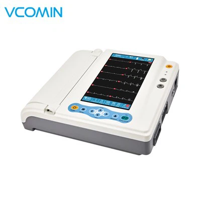
At the end of the day, a 6-channel ECG machine’s job isn’t just to print a line on a paper. It’s to turn the invisible, chaotic electrical activity of a heart into something that a clinician can read, understand, and use to help a patient. Whether it’s catching an early case of AFib, identifying a run of VPCs, or flagging VT before it turns into VF, the machine is the first step in care. If your clinic is in the market for a reliable, easy-to-use 6-channel ECG machine that’s built for the day-to-day work of seeing patients, I’m here to help. We’ve helped hundreds of clinics upgrade their ECG equipment, and I can walk you through the right model for your space, budget, and patient population, no confusing jargon, no pressure. Reach out to connect and talk through your needs—let’s make sure you have a machine that can handle every rhythm that walks through your door.
Handheld Fetal Doppler References:
Goldberger, A. L. (2018). Clinical Electrocardiography: A Simplified Approach. Elsevier.
National Heart, Lung, and Blood Institute. (2020). Atrial Fibrillation: What You Need to Know.
American College of Cardiology. (2019). ECG Interpretation for Primary Care Providers.
Zipes, D. P., & Jalife, J. (2017). Cardiac Electrophysiology: From Cell to Bedside. Elsevier.
Vcomin Technology Limited
Vcomin Technology Limited is one of the most professional 6 channel ecg machine manufacturers and suppliers in China, specialized in providing high quality customized service and OEM&ODM service. Please feel free to wholesale discount 6 channel ecg machine in stock here from our factory.
Address: 3 Fl., No.1, Kangzheng Rd., Buji Town, Shenzhen, 518110 P.R. China
E-mail: export@vcomin.com
WebSite: https://www.vcomindoppler.com/
